

1. Sign up and apply to join the challenge on the Synapse website.
2.
Submit your team information here.
Download data here.
Participants are expected to train models in their
local computational environments and to submit docker containers on Synapse
platform. A leaderboard will be maintained on the Synapse
platform during the validation phase.
We provide the code to facilitate the use of the data we release
at https://github.com/CmrxRecon/CMRxRecon2024.
A
brief description of the provided package is as follows:
- CMRxReconDemo: contains parallel imaging
reconstruction code
- ChallengeDataFormat: explains the challenge data and
the rules for data submission
- CMRxReconMaskGeneration: contains code for varied
undersampling mask generation in Task 1&2
- Evaluation: contains image quality evaluation code for
validation and testing
- Submission: contains the structure for challenge
submission
Validation of the received docker on unseen test set will be
performed on a cloud server with a configuration as follows:
You are free to use and/or refer to the CMRxRecon2024 challenge and
datasets in your own research after the embargo period (Dec 2024), provided that you cite the following
manuscripts:
Reference of the imaging acquisition protocol:
1. Wang C, Lyu J, Wang S, et al. CMRxRecon: An open cardiac MRI dataset for the competition of accelerated
image reconstruction[J]. arXiv preprint arXiv:2309.10836, 2023.
2. Wang C, Li Y, Lv J, et al. Recommendation for Cardiac Magnetic Resonance Imaging-Based Phenotypic Study:
Imaging Part. Phenomics. 2021, 1(4): 151-170.https://doi.org/10.1007/s43657-021-00018-x
Other reference (optional for citation):
1. Wang C, Jang J, Neisius U, et al. Black blood myocardial T2 mapping. Magnetic resonance in medicine.
2019,
81(1): 153-166. https://doi.org/10.1002/mrm.27360
2. Lyu J, Li G, Wang C, et al. Region-focused multi-view transformer-based generative adversarial network
for
cardiac cine MRI reconstruction[J]. Medical Image Analysis, 2023:
102760. https://doi.org/10.1016/j.media.2023.102760
3. Qin C, Schlemper J, Caballero J, et al. Convolutional recurrent neural networks for dynamic MR image
reconstruction. IEEE transactions on medical imaging, 2018, 38(1):
280-290. https://doi.org/10.1109/TMI.2018.2863670
4. Qin C, Duan J, Hammernik K, et al. Complementary time-frequency domain networks for dynamic parallel MR
image reconstruction. Magnetic Resonance in Medicine, 2021, 86(6):
3274-3291. https://doi.org/10.1002/mrm.28917
5. Lyu J, Tong X, Wang C. Parallel Imaging With a Combination of SENSE and Generative Adversarial Networks
(GAN). Quantitative Imaging in Medicine and Surgery. 2020, 10(12):
2260-2273. https://doi.org/10.21037/qims-20-518
6. Lyu J, Sui B, Wang C, et al. DuDoCAF: Dual-Domain Cross-Attention Fusion with Recurrent Transformer for
Fast Multi-contrast MR Imaging. International Conference on Medical Image Computing and Computer-Assisted
Intervention. Springer, Cham, 2022: 474-484.
7. Wang S, Qin C, Wang C, et al. The Extreme Cardiac MRI Analysis Challenge under Respiratory Motion
(CMRxMotion). arXiv preprint arXiv:2210.06385, 2022.
8. Shangqi Gao, Hangqi Zhou, Yibo Gao, Xiahai Zhuang. BayeSeg: Bayesian Modeling for Medical Image
Segmentation with Interpretable Generalizability. Medical Image Analysis Volume 89, 102889, 2023
(Elsevier-MedIA 1st Prize & MICCAl Best Paper Award 2023)
9. Wang Z, Qian C, Guo D, Sun H, Li R, Zhao B, Qu X, One-dimensional Deep Low-rank and Sparse Network for
Accelerated MRI, IEEE Transactions on Medical Imaging, 42: 79-90, 2023.
https://doi.org/10.1109/TMI.2022.3203312
10. Wang Z, et al., Deep Separable Spatiotemporal Learning for Fast Dynamic Cardiac MRI, arXiv preprint
arXiv:2402.15939, 2024. https://arxiv.org/abs/2402.15939
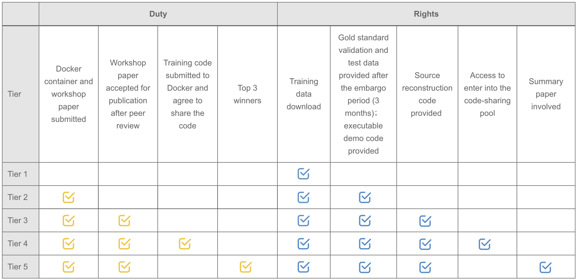
Note: Participants are not required to upload the complete training code. But teams willing to upload the original training code will be automatically entered into the code-sharing pool.
We will provide monetary awards for the top 5 winners of each
task. The prize pool is exclusively sponsored by Philips.
Task 1: Multi-contrast CMR reconstruction
| Task | Winner | Monetary Awards | Certificate | Oral Presentation | Summary Paper Involved |
| Task 1 | Top 1 | $1,000 |  |
|
|
| Task 1 | Top 2 | $500 | |
|
|
| Task 1 | Top 3 | $300 | |
|
|
| Task 1 | Top 4 | $200 | |
||
| Task 1 | Top 5 | $100 | |
Task 2: Random sampling CMR reconstruction
| Task | Winner | Monetary Awards | Certificate | Oral Presentation | Summary Paper Involved |
| Task 2 | Top 1 | $1,000 | |
|
|
| Task 2 | Top 2 | $500 | |
|
|
| Task 2 | Top 3 | $300 | |
|
|
| Task 2 | Top 4 | $200 | |
||
| Task 2 | Top 5 | $100 | |
All submissions will be reported in the leaderboard. Each participating team can participate in both
tasks. However, we only present the higher reward among the two
tasks to each team.
Prize-winning methods will be announced publicly as part of a scientific session at the MICCAI annual
meeting.
created with
Website Builder Software .