Multi-contrast CMR imaging, which involves acquiring
multiple imaging sequences with different contrast weightings, provides
valuable information for comprehensive cardiac structural and functional assessment.
However, the acquisition of multiple contrast-weighted images significantly
increases the scan time, leading to longer patient discomfort and greater
susceptibility to motion artifacts. Therefore, to reduce image
acquisition
time, there is a growing need for data-efficient and reliable reconstruction methods
to enable accelerated and high-quality multi-contrast CMR imaging.
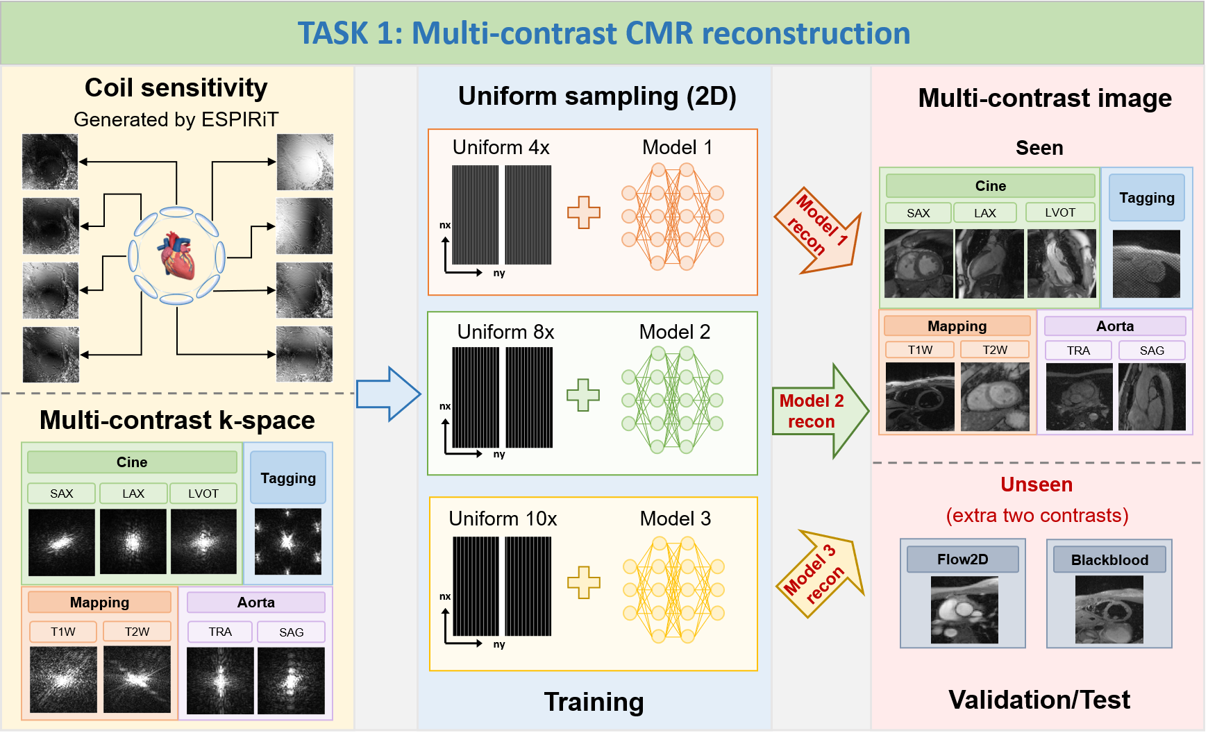
The goal of this challenge is to develop a
contrast-universal model that can 1) provide high-quality
image
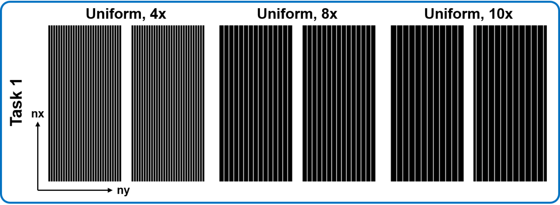
reconstruction for highly-accelerated uniform undersampling (acceleration factors are 4x, 8x and 10x, ACS not
included for calculations); 2) being able to process
multiple contrast reconstructions with different sequences,
views, and scanning protocols using a single universal model. The proposed method is supposed to offer a unified
framework that can handle various imaging contrasts, allowing for faster and more robust reconstructions across
different CMR protocols.
Note: In TASK 1, participants are allowed
to train three individual contrast-universal models to
respectively reconstruct multi-contrast data at the
aforementioned three acceleration factors; TrainingSet
includes Cine, Aorta, Mapping, and Tagging;
ValidationSet and TestSet include Cine, Aorta, Mapping,
Tagging, and other two unseen contrasts (Flow2d and
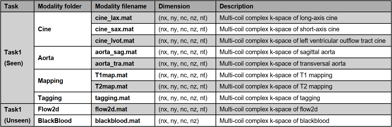
BlackBlood); the data size of Cine, Aorta, Mapping, Tagging, and Flow2d is 5D (nx,ny,nc,nz,nt); the
data
size of BlackBlood is 4D (nx,ny,nc,nz); the size of all
undersampling masks is 2D (nx,ny), the central 16
lines (ny) are always fully sampled to be used as autocalibration signals (ACS).
1) Scanner:
Siemens 3T MRI scanner (MAGNETOM Vida)
2) Image acquisition: We follow the recommendations of
CMR exams reported in the previous publication (doi: 10.1007/s43657-02100018x, 10.1007/s43657-021-00018-x).
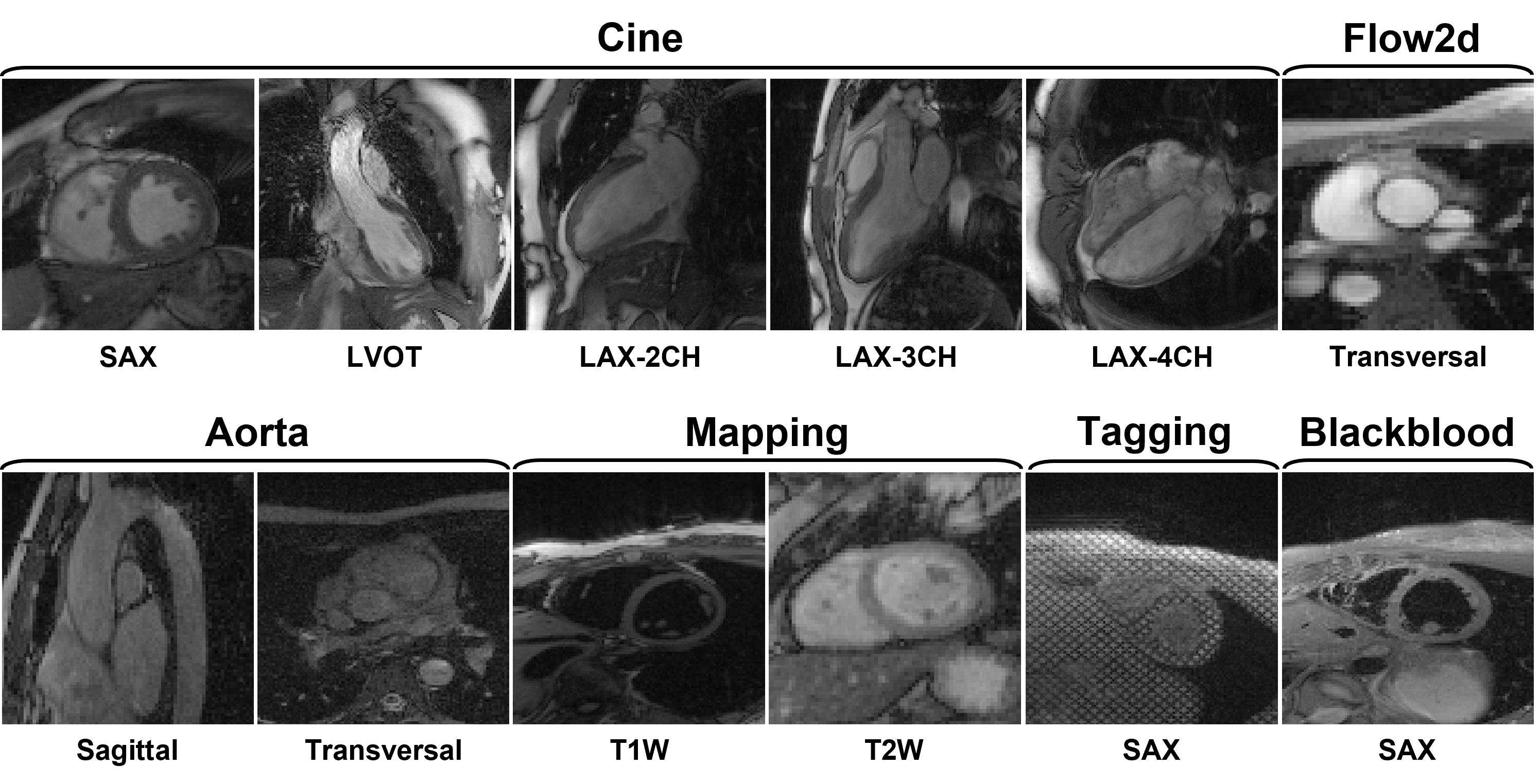
3) Dataset overview: The dataset will include multi-contrast
k-space data, consisting of cardiac cine, T1/T2 mapping, tagging, phase-contrast (i.e., flow2d), and dark-blood
imaging. It also includes imaging of different anatomical views like long-axis (LAX, including 2-chamber,
3-chamber, and 4-chamber), short-axis (SAX), left ventricul aroutflow tract (LVOT), and aortic (transversal and
sagittal views).
4) Scan protocol: We use 'TrueFISP' sequence for cine,
phase-constrast (i.e., flow2d), and tagging, and 'FLASH' sequence for T1/T2 mapping and dark-blood imaging. For
T1/T2 mapping, signals are collected at the end of the diastole with ECG triggering. Typically, 5~15 slices are
acquired for each contrast. The cardiac cycle is segmented into 12~25 phases with a temporal resolution of
around 50 ms. Typical geometrical parameters include: spatial resolution 1.5×1.5 mm2, slice thickness 8.0 mm,
and slice gap 4.0 mm.
5) Pre-processing:The raw k-space data exported from the
scanner will be processed and transformed to '.mat' format using the script provided by our vendor. A readme
file will be provided to describe the content and usage of the data.
Details of data types in Task1
Details of mask types in Task1

Metrics:
PSNR,
SSIM and NMSE
between reconstructed images and ground truth images (fully sampled data).
Ranking methods:
During the testing
and ranking phase, we will invite three radiologists to independently score the
top five teams ranked by SSIM. The scoring will cover three
aspects: image
quality, image artifacts, and clinical utility. We will consider both the
radiologists' scores and the SSIM results to generate a comprehensive ranking.
Participating
teams are required to submit docker containers and process all the cases in the
test set on our server. For the cases without valid output, we will assign it
to the lowest value of metric.
1) To ensure the fairness of this challenge, you are only allowed to use the datasets provided by fastMRI,
CMRxRecon2023, and CMRxRecon2024. Data augmentation based on the training dataset is allowed.
2) In TASK 1, participants are allowed to train three
individual contrast-universal models to respectively reconstruct multi-contrast data at three
acceleration factors.
created with
Website Builder Software .